
Are You At Risk For Macular Degeneration?

Can you believe it’s already February? A new month means a new opportunity to take better care of your eyes. And since February is officially AMD Awareness month, the timing couldn’t be better. If you have no idea what AMD is - don’t worry - the EyeQ blog has you covered. Much like our travels into Glaucoma Awareness for January, this month will guide you through the things you need to know about age related macular degeneration aka AMD. So keep reading below where we will start with what this disease is; and be sure check back in regularly for even more information throughout the month.
What is AMD?
To better understand AMD, let’s first start with a little eye anatomy. The entire inside of the eye is lined with a thin layer of tissue called the retina. The retina contains light sensitive photoreceptor cells that are responsible for sending impulses to the brain - via the optic nerve - that are then translated into the images we see.
Located near the center of the retina at the very back of the eye is the macula - the part of the eye responsible for detailed central vision. Although the macula is small, it is a lot more sensitive than the surrounding areas of the retina. It is, after all, what we rely on for the vision we use on a daily basis for tasks such as reading ,driving, recognizing faces, etc. The area of retina surrounding the macula is more specifically known as the peripheral retina. With the macula in charge of central vision, the peripheral retina controls what we see out to the sides - our peripheral vision.
Age related macular degeneration is a progressive eye disease that causes a breakdown or degeneration of the macula. This can result in vision loss ranging from a decrease in color and light perception to complete loss of central vision - leaving only a black spot in the middle of anything that is viewed. Peripheral vision (aka side vision) will stay in tact since AMD only affects the macula and not the peripheral retina.
So why and how does this happen? There are actually two different forms of AMD - wet/neovascular and dry/atrophic. Let’s take a look.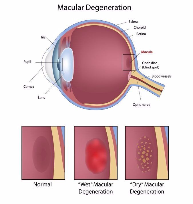
Dry AMD
Dry macular degeneration, also referred to as atrophic macular degeneration, is actually the more common form of AMD. In fact, according to the Macular Degeneration Partnership , 90% of people who have macular degeneration have the “dry” form.
Remember that layer of light sensitive cells we mentioned earlier? Below those lies another layer of cells called retinal pigment epithelial cells (RPE). These cells are responsible for supporting the photoreceptors by providing them with nutrients and waste removal. In the dry form of AMD, this layer of RPE cells begins to degenerate - usually as part of the eye’s natural aging process. This death of cells is also called atrophy - which is why dry AMD is referred to as atrophic AMD.
Typically the visual damage associated with dry AMD is not quite as severe or rapid as the other form of AMD - wet. Those who experience the dry form of AMD have a good chance of maintaining central vision of 20/40 or better, but will experience limitations when reading and under dim lighting conditions. However, if left untreated, permanent vision loss will occur.
Wet AMD
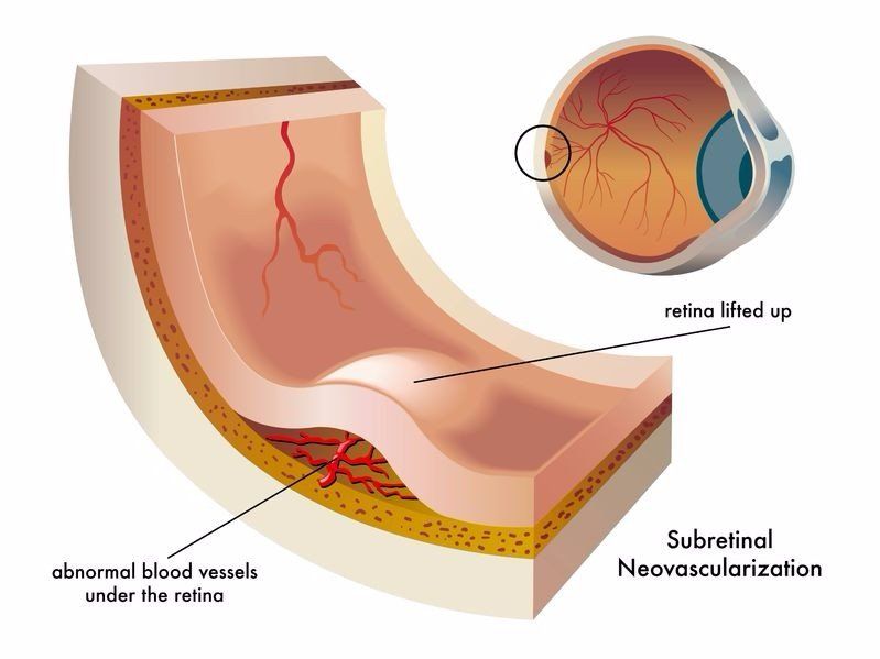
Wet macular degeneration is the more severe form of AMD. With this type, there is a lack of adequate oxygen supply to the macula that in turn causes new and abnormal blood vessels to grow in an attempt to compensate for the deficiency.
These vessels will grow through the membrane behind the retina, much like the roots of a tree will sometimes grow through the cracks in a sidewalk. Unfortunately the vessels that grow are usually very fragile and have a tendency to leak fluid or blood - called exudate - which in turn results in damage that causes blurred and distorted vision as well as rapid central vision loss.
The term exudate is why wet AMD is also known as exudative macular degeneration. Also, if you’ve wondered why these two are referred to as wet and dry - the presence of fluid in the wet form vs the absence of fluid or blood in the dry form is what is being referenced.
Visual disturbances from the wet form of AMD are usually more noticeable and progress faster than that from dry AMD. In fact, although wet macular degeneration only accounts for around 10% of AMD cases, 90% of AMD related blindness is caused by the wet form.
A Side Note About Drusen...
Before we get into what causes macular degeneration, let’s take a look at an important component of both wet and dry AMD called drusen. Drusen are small yellow deposits that are made up of fatty proteins called lipids. These appear in a layer of the retina called Bruch’s membrane and may be the result of a decrease in the eye’s ability to dispose of waste products when the RPE layer begins to deteriorate.
There are two different types of drusen with varying levels of risk. For example, drusen can be hard and small with distinct edges. These are usually spaced far apart from each other and might not cause vision problems for a long time - if ever.
Drusen can also become softer, larger, and cluster closer together with less defined edges. This type of drusen actually increases your risk for wet macular degeneration.
Having drusen doesn’t always mean you have macular degeneration, but it can be an early sign of the disease. If your doctor mentions seeing drusen during your exam, be sure to have regular eye health examinations to watch for any changes.
What Causes AMD?
The exact cause of the breakdown of cells that leads to dry AMD is uncertain. However, it is thought to be mainly attributed to the eye’s natural aging process. Much like the rest of the body breaks down over time - as does the eye. There are a certain number of things that can cause this to happen faster or even prematurely, but we will get to that a little later.
When it comes to the cause of wet AMD, everyone who has the wet form started out with dry AMD even if they were unaware of it. According to the Macular Degeneration Partnership, scientists currently theorize that when the body recognizes the disruption of circulation in the eye caused by the degeneration of dry AMD, a growth factor called VEGF (vascular endothelial growth factor) is triggered. This is what causes the growth of new blood vessels. VEGF can be a positive thing in certain scenarios such as sending new blood vessels to the part of the heart affected by a heart attack. However, VEGF causes a negative impact when it directs them to a cancerous tumor causing it to grow - or in the case of the eye with wet AMD.
Know Your Risk Factors
There is currently no cure for macular degeneration, and while there are effective treatment options available, the best method is one of prevention. Knowing your risk factors, your family history, and keeping regular eye health appointments are all an important part of making sure your vision lasts as long as you do. Here are the American Academy of Ophthalmology’s top 5 AMD risk factors:
- Having a family history of AMD
- Being obese or leading a sedentary lifestyle
- Smoking
- Being over the age of 60
- Having high blood pressure
Be your own Valentine this year and treat your eyes to an exam! Also stay tuned for AMD Awareness parts 2 &3 where we will go over symptoms and treatment options.